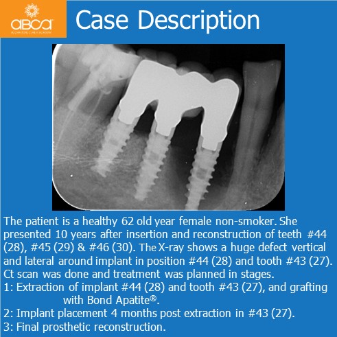
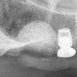
The patient is a healthy 62 old year female non-smoker. She presented 10 years after insertion and reconstruction of teeth #44 (28), #45 (29) & #46 (30). The X-ray shows a huge defect vertical and lateral around implant in position #44 (28) and tooth #43 (27).
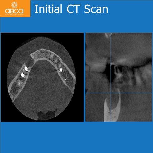
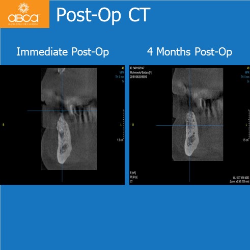
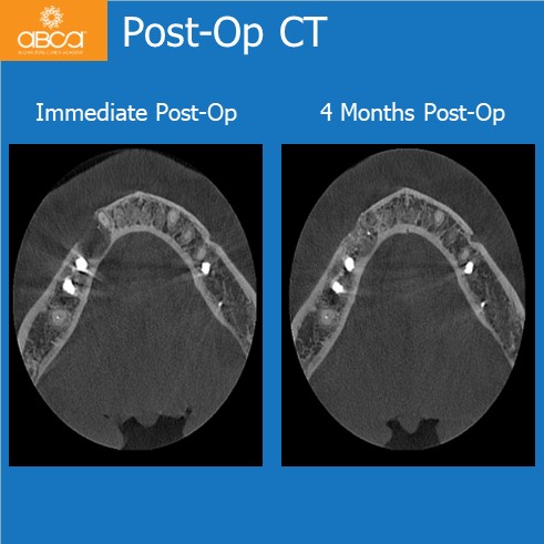
CT scan was done and treatment was planned in stages.
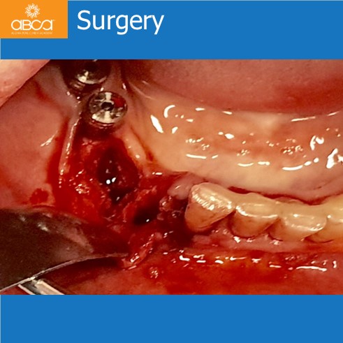
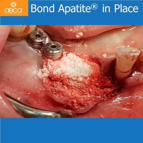
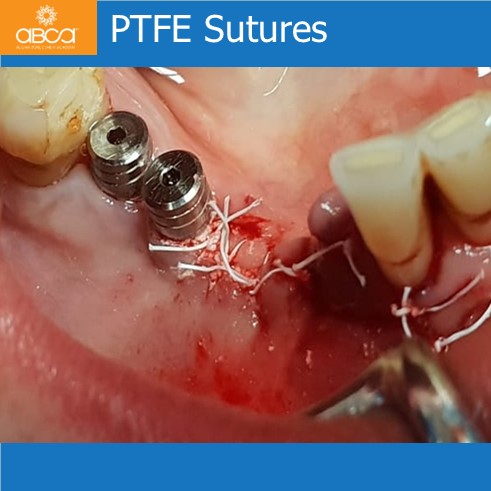
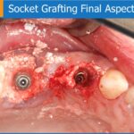
- Extraction of implant #44 (28) and tooth #43 (27), and grafting with Bond Apatite®.
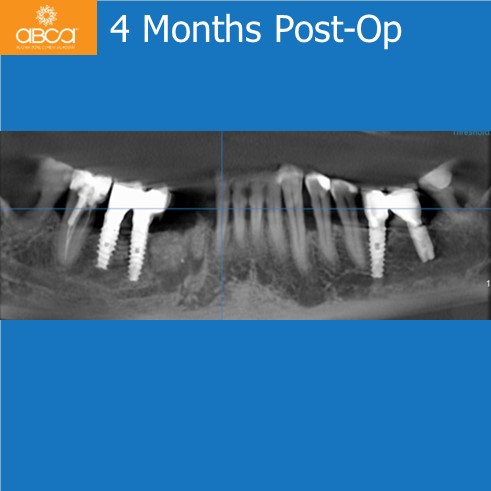
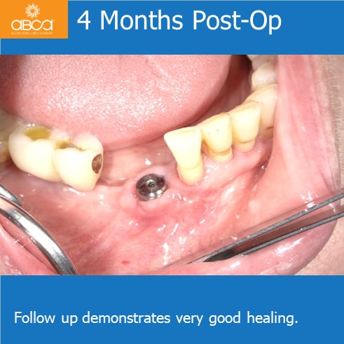
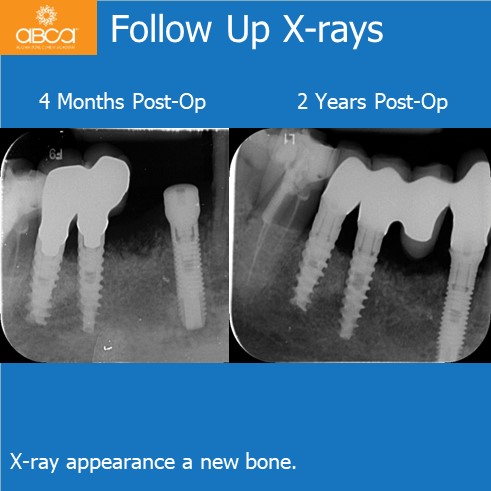
- Implant placement 4 months post extraction in #43 (27).
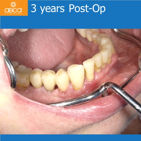
- Final prosthetic reconstruction.
![](https://www.augmabio.com/wp-content/uploads/2024/05/ewagbr4-1.jpg)
![](https://www.augmabio.com/wp-content/uploads/2024/05/ewagbr4-2.jpg)
![](https://www.augmabio.com/wp-content/uploads/2024/05/ewagbr4-3.jpg)
![](https://www.augmabio.com/wp-content/uploads/2024/05/ewagbr4-4.jpg)
![](https://www.augmabio.com/wp-content/uploads/2024/05/ewagbr4-5.jpg)
![](https://www.augmabio.com/wp-content/uploads/2024/05/ewagbr4-6.jpg)
![](https://www.augmabio.com/wp-content/uploads/2024/05/ewagbr4-7.jpg)
![](https://www.augmabio.com/wp-content/uploads/2024/05/ewagbr4-8.jpg)
![](https://www.augmabio.com/wp-content/uploads/2024/05/ewagbr4-9.jpg)
![](https://www.augmabio.com/wp-content/uploads/2024/05/ewagbr4-10.jpg)
![](https://www.augmabio.com/wp-content/uploads/2024/05/ewagbr4-11.jpg)
![](https://www.augmabio.com/wp-content/uploads/2024/05/ewagbr4-12.jpg)
![](https://www.augmabio.com/wp-content/uploads/2024/05/ewagbr4-13.jpg)