The patient is a healthy, 66-year-old female. The root of tooth #13 (6) is broken. In the area of #16 (3) only 1.3 mm of bone is present and there is no possibility of prosthetic restoration. She presents with pain and discomfort.
Treatment plan consists of 3 steps.
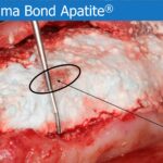
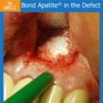
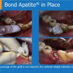
Step 1: Extraction of tooth #13 (6) and filling the socket with Bond Apatite®. 7 days post-op the wound opened, however most of the cement stayed in place and the wound closed on its own within two weeks without bacterial contamination.
Step 2: 4 months post-op Augma Lift™ in the area of #16 (3) and implant placement in #13 (6) and #14 (5). Good control of the Schneiderian membrane allows for safe elevation. 1cc Bond Apatite® fills the sinus cavity.
Step 3: A histology sample (bone shaving) taken from the area of #16 (3) show a lot of living new bone. Microphotography shows points with a smooth transition between hydroxyapatite crystals and new bone. There is no observable inflammation at this time.
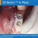
4 months later implant #16 (3) is opened with good osteointegration. Histology, X-ray and clinical view confirms more than 7mm of new bone from Augma Lift™. Conclusion is that Augma Lift™ is a safe, easy and innovative alternative to the classic sinus lift procedure.
CLINICAL CASE Augma Lift™ and Implants in 3 Steps Dr. Damian Dudek DMD, PhD, PolandCase Description – The patient is a healthy, 66-year-old female. The root of tooth #13 (6) is broken. In the area of #16 (3) only 1.3 mm of bone is present and there is no possibility of prosthetic restoration. She presents with pain and discomfort. Treatment plan consists of 3 steps. Step 1: Extraction of tooth #13 (6) and filling the socket with Bond Apatite ® . 7 days post-op the wound opened, however most of the cement stayed in place and the wound closed on its own within two weeks without bacterial contamination. Step 2: 4 months post-op Augma Lift™ in the area of #16 (3) and implant placement in #13 (6) and #14 (5). Good control of the Schneiderian membrane allows for safe elevation. 1cc Bond Apatite ® fills the sinus cavity. Step 3: A histology sample (bone shaving) taken from the area of #16 (3) show a lot of living new bone. Microphotography shows points with a smooth transition between hydroxyapatite crystals and new bone. There is no observable inflammation at this time. 4 months later implant #16 (3) is opened with good osteointegration. Histology, X-ray and clinical view confirms more than 7mm of new bone from Augma Lift™. Conclusion is that Augma Lift™ is a safe, easy and innovative alternative to the classic sinus lift procedure.Pre-Op PanoramicAdvanced InflammationPre-Op 1.3mm of Bone – In area of #16 (3) there is only 1.3 mm of cortical bone.#13 (6) – 7 Days Post-Op – Step 1: 7 days post extraction and augmentation of tooth #13 (6).Augma Lift™ Technique – Step 2: Augma Lift™ technique. The bone plate is visible inside the trepanation.Elevating the Membrane – Elevating the sinus membrane with Augma Lift™ osteotome.Implants #13(6) & #14(5) – Step 2: Implants in #13 (6) and #14 (5), 4 months post augmentation.Implants & Bond Apatite® – Immediate post-op X-ray of implants #13(6) & #14(5). Bond Apatite® seen in the area of #16(3) after augmentation with Augma Lift™.7 Days Post-Op – Seven days after augmentation, Bond Apatite® is visible and the sinus elevation is measured nearly 10 mm.Orbital CBCT7 Days Post-OpFlap Opening #16(3) – Step 3: Implant placement in #16 (3).Implant Placement #16(3) – Bicon implant in the new bone.6 Months Post-Op – Histology showing new bone (1) and hydroxyapatite crystals (2) are visible. The next slide shows a close-up of the circled area. Focus 100x H&E staining.6 Months Post-Op – Bone shavings from #16 (3). The arrow shows the smooth transition between HPA and new bone. Focus 200x, H&E staining7mm Regenerated Bone – Regenerated bone is measured at 7mm.4 Months Post Placement – Healing abutment 4 months after implant placement.Final X-ray #16(3)