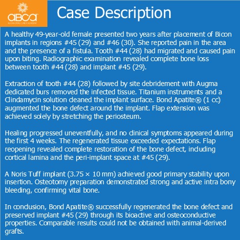
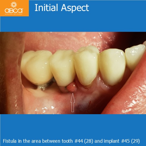
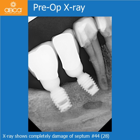
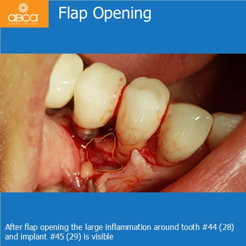
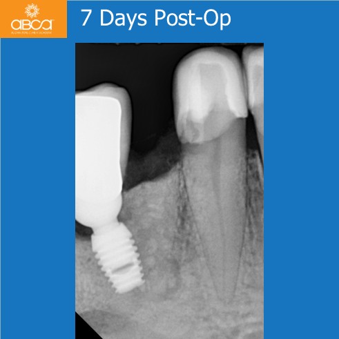
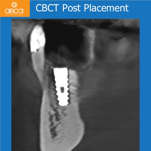
A healthy 49-year-old female presented two years after placement of Bicon implants in regions #45 (29) and #46 (30). She reported pain in the area and the presence of a fistula. Tooth #44 (28) had migrated and caused pain upon biting. Radiographic examination revealed complete bone loss between tooth #44 (28) and implant #45 (29).
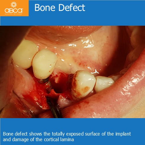
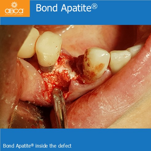
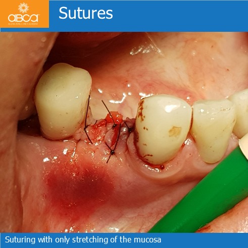
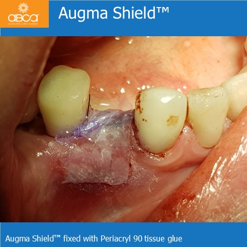
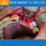
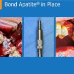
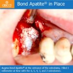
Extraction of tooth #44 (28) followed by site debridement with Augma dedicated burs removed the infected tissue. Titanium instruments and a Clindamycin solution cleaned the implant surface. Bond Apatite® (1 cc) augmented the bone defect around the implant. Flap extension was achieved solely by stretching the periosteum.
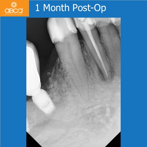
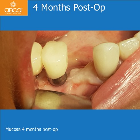
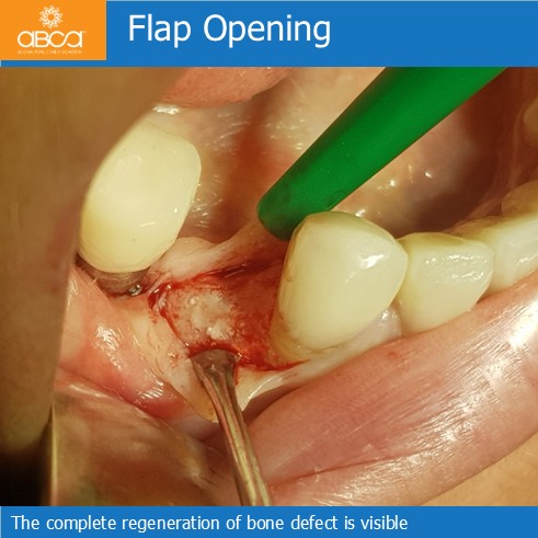
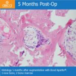
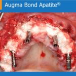
Healing progressed uneventfully, and no clinical symptoms appeared during the first 4 weeks. The regenerated tissue exceeded expectations. Flap reopening revealed complete restoration of the bone defect, including cortical lamina and the peri-implant space at #45 (29).
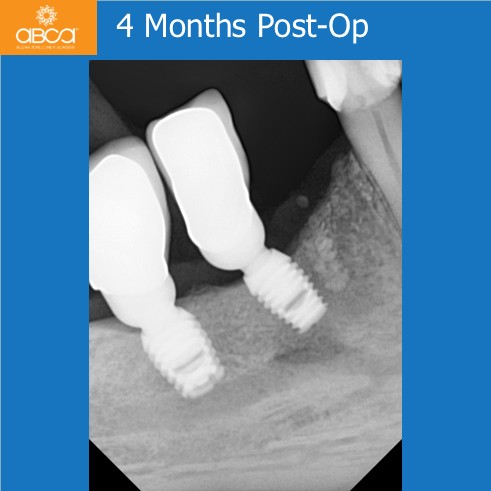
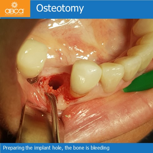
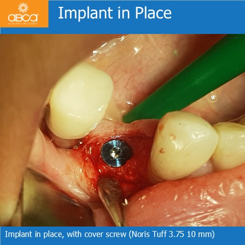
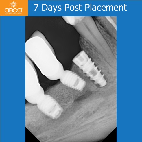
A Noris Tuff implant (3.75 × 10 mm) achieved good primary stability upon insertion. Osteotomy preparation demonstrated strong and active intra bony bleeding, confirming vital bone.
In conclusion, Bond Apatite® successfully regenerated the bone defect and preserved implant #45 (29) through its bioactive and osteoconductive properties. Comparable results could not be obtained with animal-derived grafts.