Alveolar Ridge Augmentation with Simultaneous Implant Insertion
The patient is a 46-year-old female who came to the clinic for rehabilitation of the lower jaw. The initial consultation revealed generalized chronic periodontitis which led to the loss of posterior teeth 36 (19), 37 (18), 45 (29), 46 (30) & 47 (31) , loss of tooth 35 (20) due to decay, and the presence of wisdom teeth 38 (17) and 48 (32).
The treatment plan began with Er-Yag laser assisted treatment of chronic periodontitis with the extraction of teeth 35 (20), 38 (17) and 48 (32) to stabilize the disease. This will avoid the inflammation of gingiva which can inhibit bone regeneration and lead to unpredictable gingival healing.
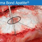
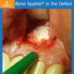
Two months after the laser treatment implants were placed at 34 (21), 35 (20), 36 (19), 45 (29), 46 (30) & 47 (31). Alveolar regeneration is performed with Bond Apatite® to increase bone width.
4-month post-op images confirm very good alveolar bone regeneration around the implants.
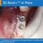
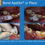
CLINICAL CASE Alveolar Ridge Augmentation with Simultaneous Implant Insertion Dr. Mirela Shveys DMD, MD, OMS, RomaniaCase Description – The patient is a 46-year-old female who came to the clinic for rehabilitation of the lower jaw. The initial consultation revealed generalized chronic periodontitis which led to the loss of posterior teeth 36 (19), 37 (18), 45 (29), 46 (30) & 47 (31) , loss of tooth 35 (20) due to decay, and the presence of wisdom teeth 38 (17) and 48 (32). The treatment plan began with Er-Yag laser assisted treatment of chronic periodontitis with the extraction of teeth 35 (20), 38 (17) and 48 (32) to stabilize the disease. This will avoid the inflammation of gingiva which can inhibit bone regeneration and lead to unpredictable gingival healing. Two months after the laser treatment implants were placed at 34 (21), 35 (20), 36 (19), 45 (29), 46 (30) & 47 (31). Alveolar regeneration is performed with Bond Apatite® to increase bone width. 4-month post-op images confirm very good alveolar bone regeneration around the implants.CBCT of the Lower Jaw – CBCT of the lower jaw reveals a socket defect after the extraction of tooth #45 (29), with a loss of vestibular cortical wall, root #35 (20) with no buccal cortex and alveolar width of 3mm on both sides of the jaw.CBCT of the Lower Jaw – CBCT of the lower jaw reveals a socket defect after the extraction of tooth #45 (29), with a loss of vestibular cortical wall, root #35 (20) with no buccal cortex and alveolar width of 3mm on both sides of the jaw.Initial Aspect – The initial aspect of area #45 (29) – #48 (32), prior to the extraction of #48 (32) and augmentation. 4mm of attached gingiva 2 months after Er-Ya laser assisted treatment of chronic periodontitis.Alveolar Crest 3mm Width – The alveolar crest with a 3 mm width on the upper part.Implants in Place – Implants placed in #45 (29), #46 (30) and #47 (31) and the extraction of tooth #48 (32). The buccal cortex over the implants is under 1mm. The horizontal defect on the #45 (20) implant is 2mm. Round bur holes are made deep into the cancellous bone to obtain bleeding.Bond Apatite® in Place – Bond Apatite® is placed over the implants, horizontally, vertically and bilaterally.SuturesBone Defect at #35 (20) – Vertical post-extract bone defect of 4 mm at #35 (20). Round bur holes are made deep into the cancellous bone to obtain bleeding.Implants #35(20) – #37(18) – Implants are placed in #35 (20), #36 (19) and #37 (18). The buccal cortex over the implants is under 1mm. The horizontal defect on the #35 (20) implant is 4mm.Bond Apatite® in Place – Bond Apatite® is placed over the implants, horizontally, vertically and bilaterally.4 Months Post-Op – Lower, right mandible: implant exposure 4 months post-op.Implant Exposure – Lower, right mandible: 4 months post-op the implants were exposed. The buccal cortical wall was regenerated at area #34 (21) and #44 (28). The alveolar width is 6mm.Implant Exposure – Lower, right mandible: Careful management of gingiva lead to a keratinized collar of 2mm around the healing cap.4 Months Post-Op – Lower, left mandible: Implant exposure 4 months post-op.Implant Exposure – Lower, left mandible, implant exposure.Implant Exposure – Lower, left mandible: Careful management of gingiva lead to a keratinized collar of 2mm around the healing cap.CBCT #35 (20) – #45 (29) – CBCT post augmentation confirms very good alveolar bone regeneration around the implants.CBCT #36 (19) – #46 (30) – CBCT post augmentation confirms very good alveolar bone regeneration around the implants.CBCT #37 (18) – #47 (31) – CBCT post augmentation confirms very good alveolar bone regeneration around the implants.