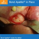
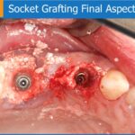
This case demonstrates the clinical and biological versatility of biphasic calcium sulphate. All digital. The last canine remaining was used for vertical dimension of occlusion. No angled MUA for the original implant is available, so the implant was removed at time of surgery.
The removal tool fractured and so it was decided to leave the implant. Implants were place and were scanned again, then extracted and the lower right canine socket was grafted.
The lab failed to deliver provisional later that day so the patient’s tongue damaged the flap over the buried implant (sutured with chromic since it was intended to be covered).
Here is where Augma shines. There was no infection and healing was great. The temp was placed 7 days post-op, and the final was placed at 4 months post-op.

2 Comments
Was that a flowable composite that was placed on the tissue after removal of the remaining canine?
“Yes. It was flowable light cured dentin liner.” – Dr. Michael Katzap