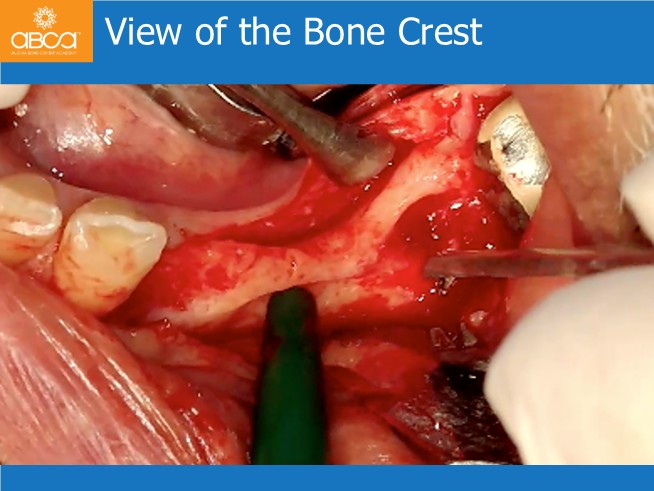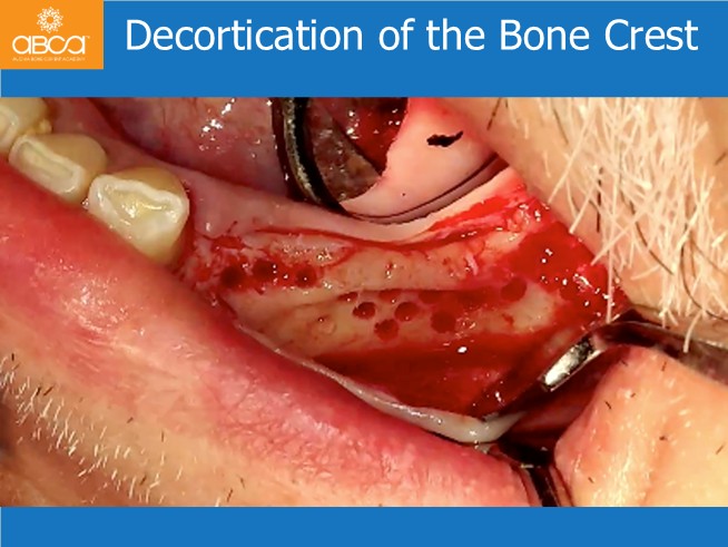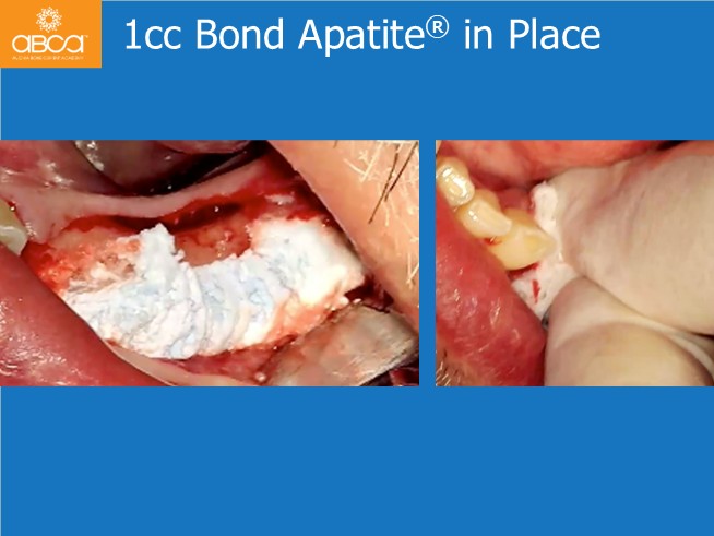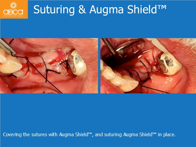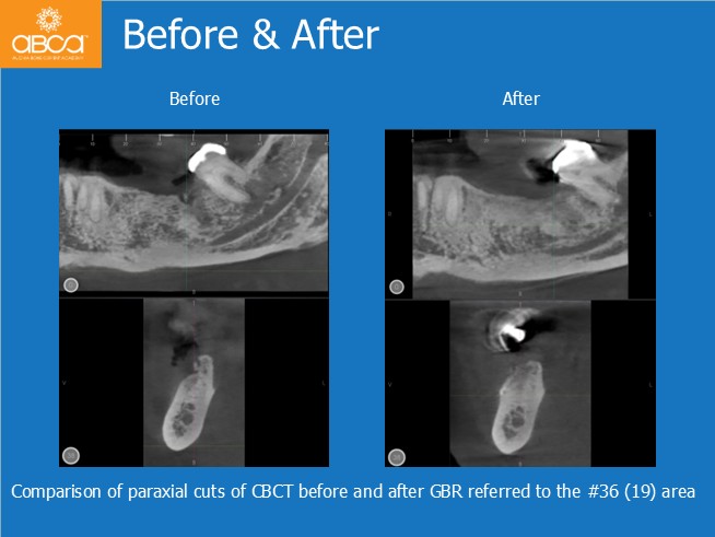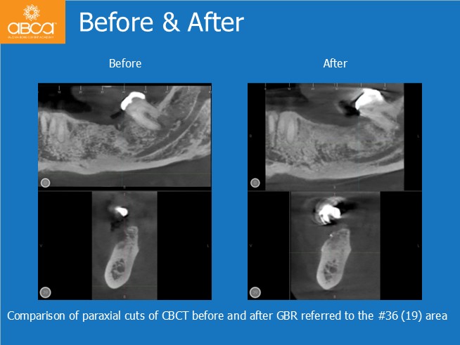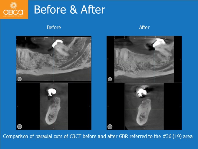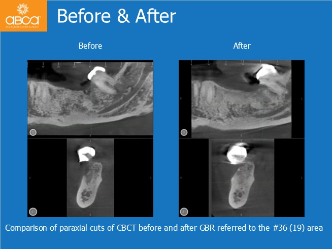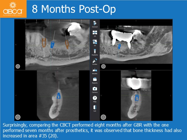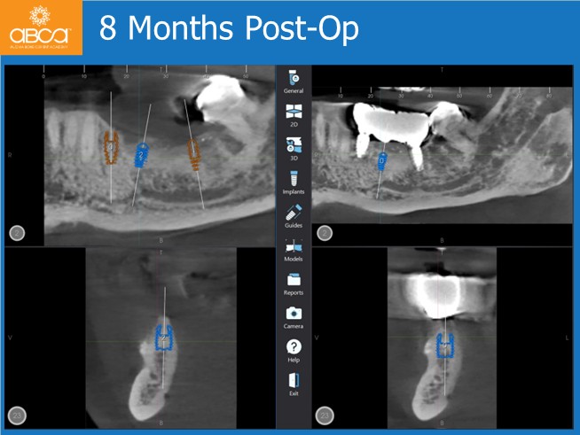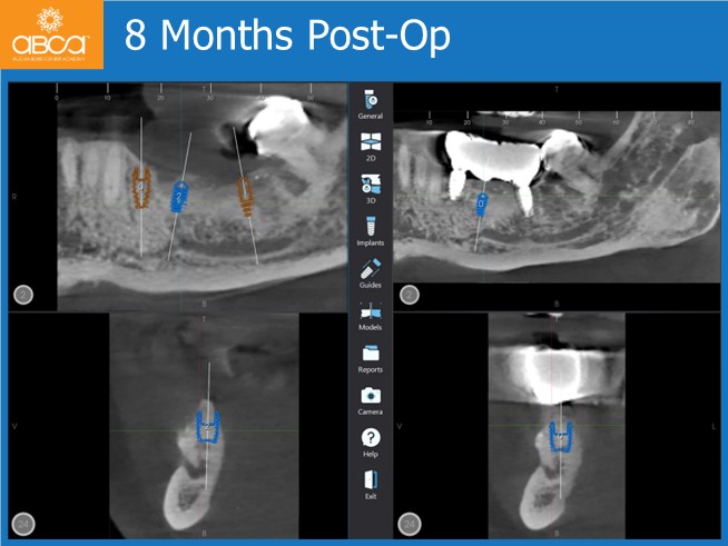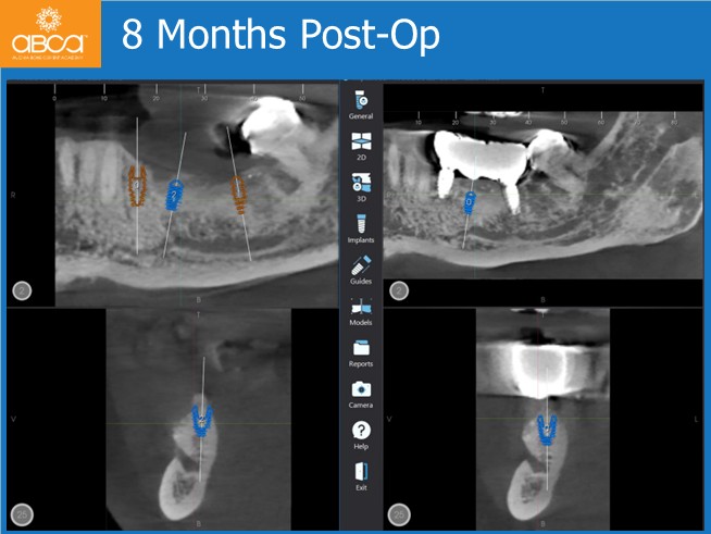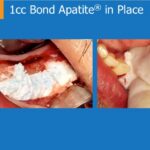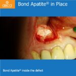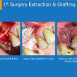
A healthy 69-year-old man in good health presented to us with a request for prosthetic rehabilitation of the distal edentulism of the third quadrant from teeth #34 (21) to #36 (19). His desire was to obtain a fixed rehabilitation.
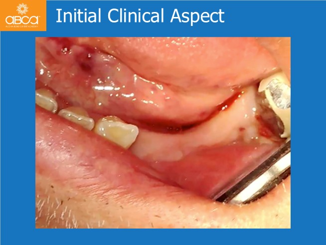
On physical examination, the patient showed edentulism of teeth #34 (21), #35 (20), and #36 (19), with a capped crown on #37 (18). Upon palpation, he exhibited a very thin, non-linear bone crest and a band of keratinized mucosa approximately 5-6 cm long in the buccal-vestibular direction.
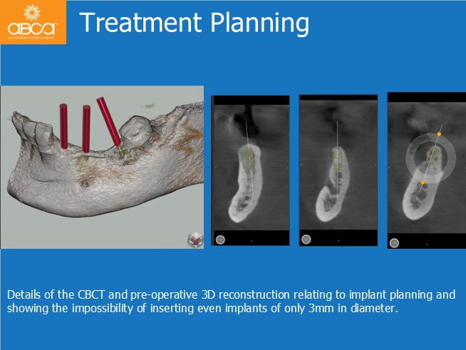
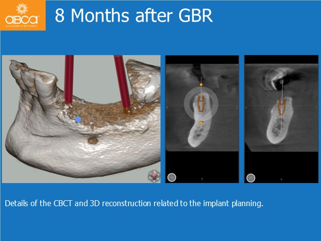
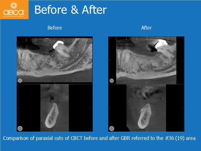
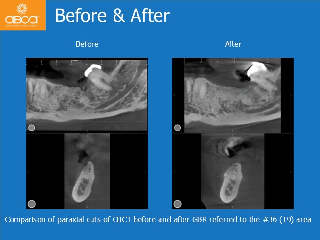
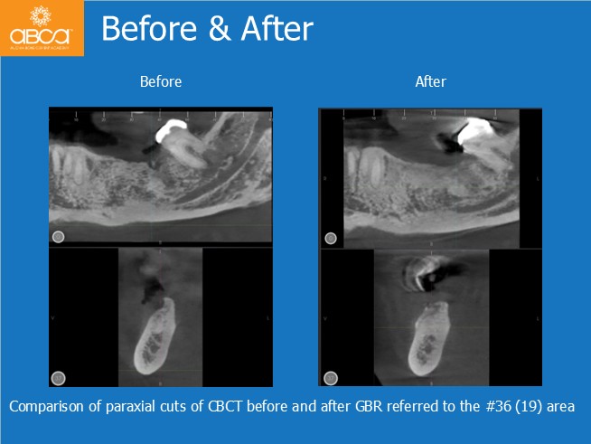
Preoperative CBCT confirmed the bone morphology, with the impossibility of placing implants in areas #35 (20) and #36 (19). It was decided to approach a lateral alveolar ridge augmentation.
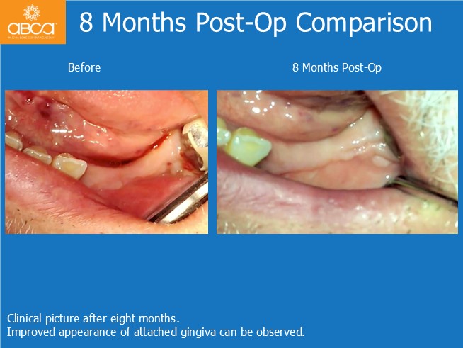
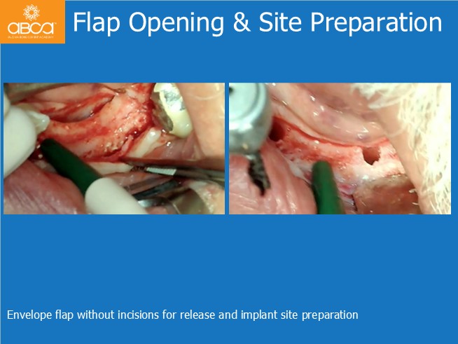
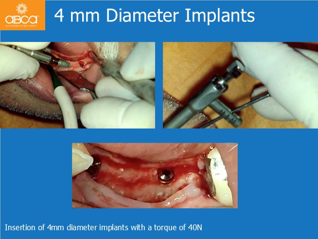
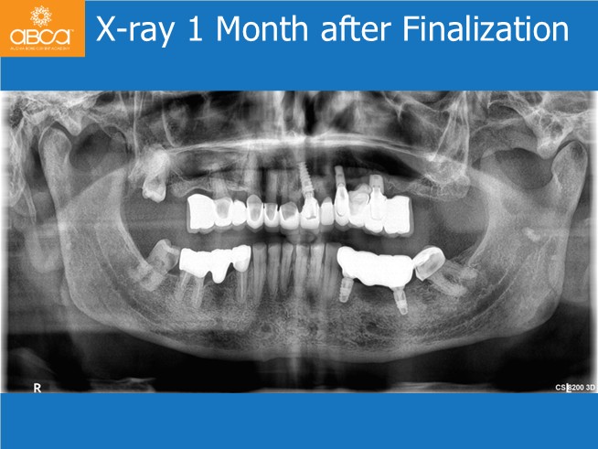
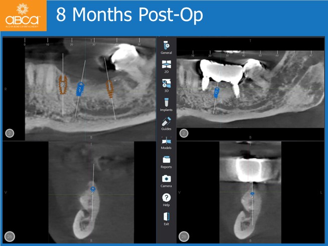
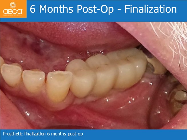
The surgical procedure was successfully completed in two stages: 1) bone cement grafting; 2) After eight months, implants were inserted in areas #34 (21) and #37 (19). Subsequently, a bridge was placed, restoring all edentulous areas, to the patient’s great satisfaction with the overall favorable postoperative outcome.
Interestingly, the radiological follow-up six months after the prosthesis placement revealed further increase in volume, potentially warranting the insertion of another implant.