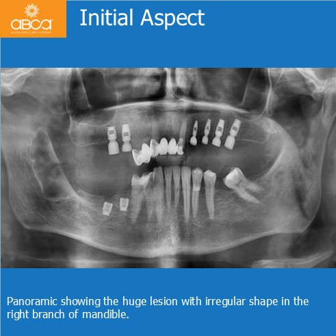
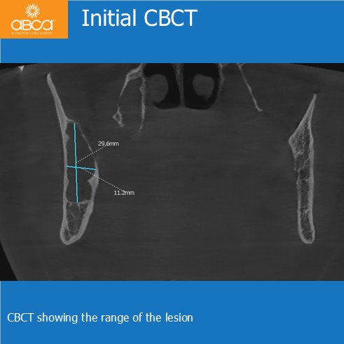
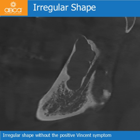
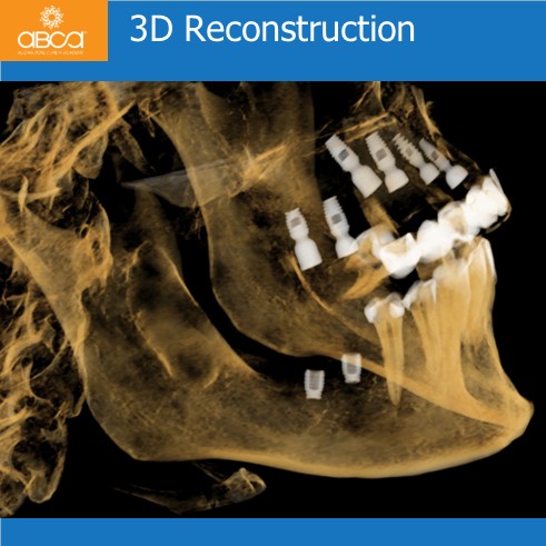
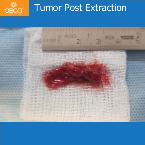
The patient is a 56-year-old male, who presents with pain and swelling in the right branch of the mandible. Radiographs and CBCT reveal an osteolytic lesion with a radiolucent appearance suggestive of a dentigerous cyst; no Vincent symptom is present. The initial sample sent for histopathological analysis confirms an odontogenic cyst.
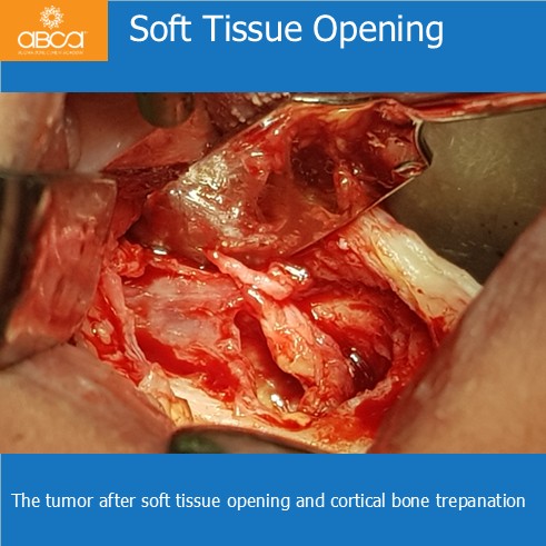
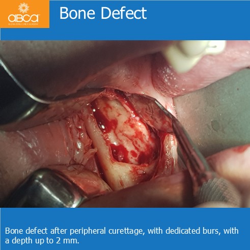
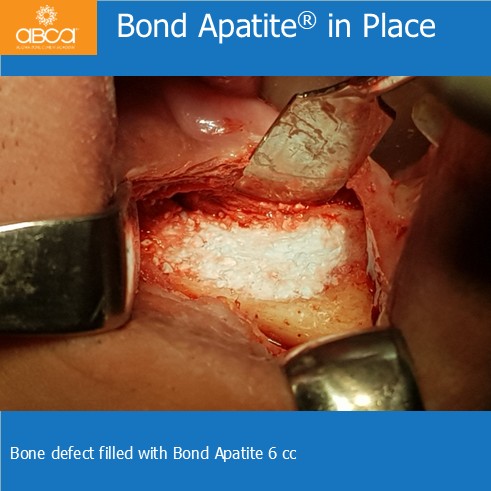
Treatment includes tumor removal with peripheral bone curettage to a depth of approximately 2 mm, using dedicated Augma degranulation burs. Dr. Dudek fills the bone defect is filled with 6 cc of Bond Apatite®, and sent the lesion for histopathological examination. The final diagnosis is a keratocystic odontogenic tumor (KCOT), parakeratotic type.
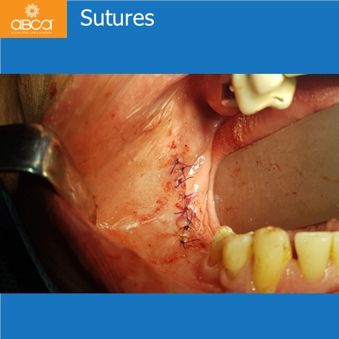
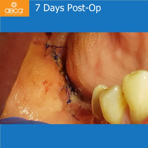
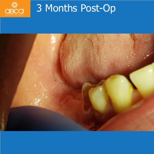
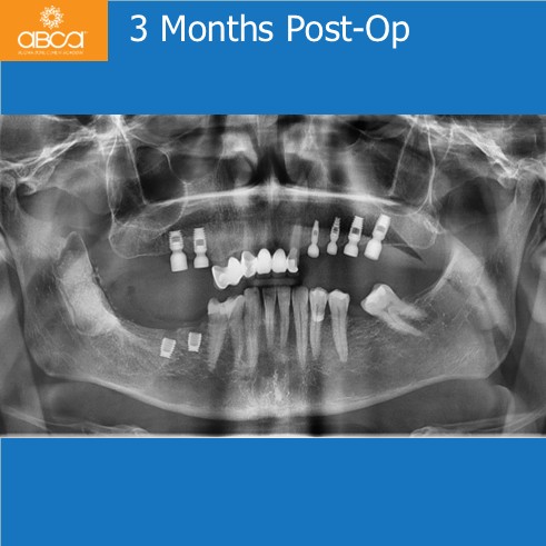
Healing progress well, with suture removal 7 days post-op. At the 3 month follow up visit there is no recurrence or secondary inflammatory symptoms observed, and the patient remains under regular monitoring.