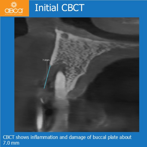
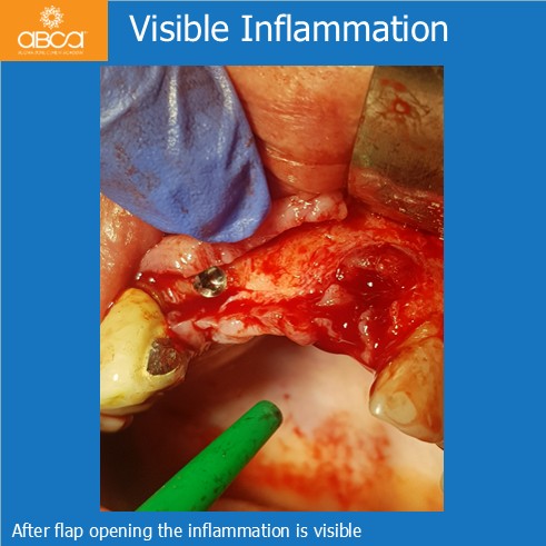
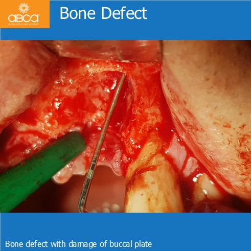
A 76-year-old male patient presented with a fractured bridge in the anterior right maxilla that was not amenable to prosthetic restoration. He reported mild pain and discomfort. Clinical and radiographic evaluation also revealed significant inflammation and a bone defect in the region of tooth #21 (9), including approximately 7.0 mm loss of the buccal cortical plate.
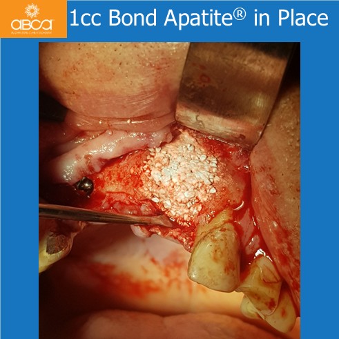
After removing the bridge, Dr. Dudek identified sufficient bone volume at site #12 (7) to place a standard screw implant (Noris Tuff 3.75 × 13 mm). At site #21 (9), only the bone defect remained, with confirmed buccal plate loss. Filling of the defect with 1 cc of Bond Apatite®.
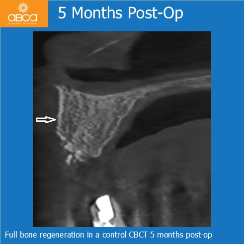
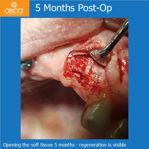
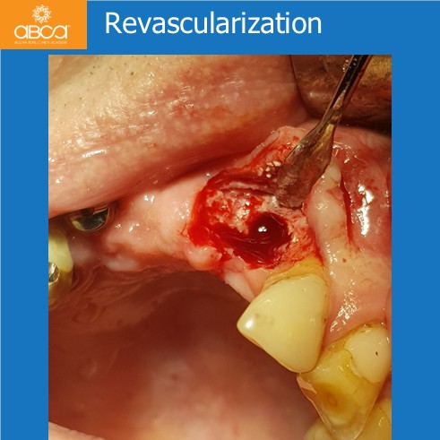
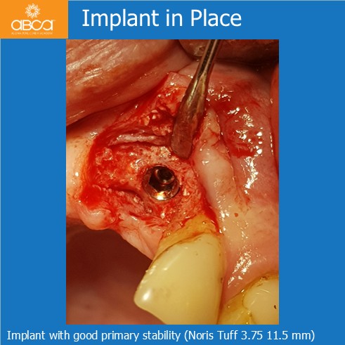
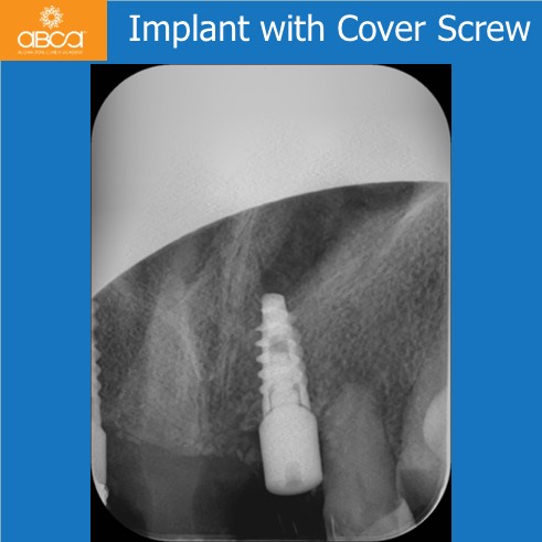
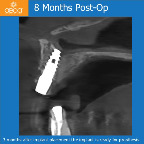
Implant placement at site #21 (9), 5 months post-op. Clinical evaluation demonstrated complete bone regeneration, including restoration of the buccal cortical plate, with excellent revascularization throughout the regenerated site. Histological analysis of a biopsy obtained before implant placement confirmed these findings. In addition, the implant at #21 (9) (Noris Tuff 3.75 × 11.5 mm) achieved excellent primary stability, allowing placement of a healing abutment. After an additional 3 months, the implant site was ready for prosthetic rehabilitation.