Augma Biomaterials

Lateral Augmentation

SOCKET
GRAFTING
LATERAL
AUGMENTATION
SINUS
LIFT
FULL ARCH
RECONSTRUCTION
AESTHETIC
ZONE
CYST
ENUCLEATION

PERI IMPLANTITIS

ZYGOMA

Lateral Augmentation

To expose the defect perform a minimal flap reflection, of no more than 3mm past the mucogingival junction or as much as needed to expose the defect. Decorticate the buccal plate and place Augma Bond Apatite®. Compact the material well with a sterile gauze. Closure of the flap is performed with tension after stretching the flap. Do not perform soft tissue manipulation to gain a tension free flap.  If primary closure is not possible then cover the exposed graft and soft tissue with Augma Shield™.


Surgical Protocol

How-To Videos

More Clinical Content on Augmaflix

Products for Lateral Augmentation

Lateral Augmentation – 2 mm ridge

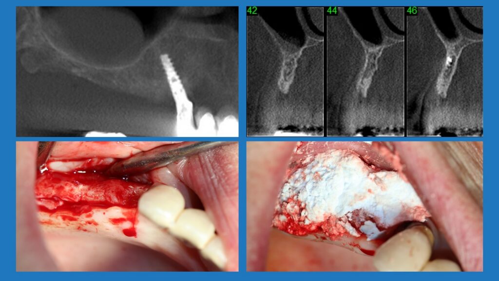
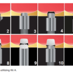
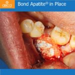
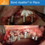
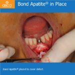
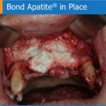
Patient in her sixties come for implant treatment in the maxilla on the right side. The ridge was very thin, about 2 mm. A lateral augmentation to thicken the ridge with Bond Apatite was performed. The implant in tooth #13 (6) is of 3 mm in diameter, the other implants #14 (5), #15 (4) and #17 (2) are of 3.75 mm in diameter. The use of the Bond Apatite in the case makes it a simple surgery, without a membrane, and with minimally invasive protocols.

[gap]

[ux_slider]

[ux_image id=”17199″ image_size=”original”]

[ux_image id=”17206″ image_size=”original”]

[/ux_slider]

Leave a Reply
Select the fields to be shown. Others will be hidden. Drag and drop to rearrange the order.
  • Image
  • SKU
  • Rating
  • Price
  • Stock
  • Availability
  • Add to cart
  • Description
  • Content
  • Weight
  • Dimensions
  • Additional information
Click outside to hide the comparison bar
Compare
Shopping cart close