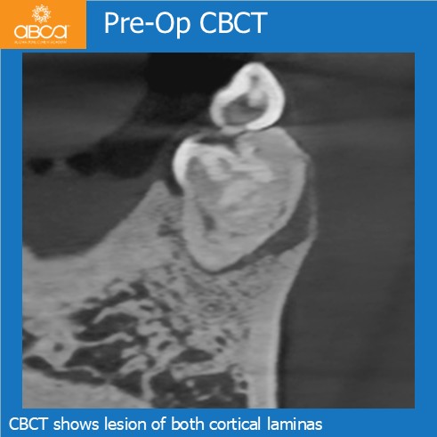
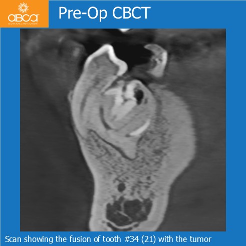
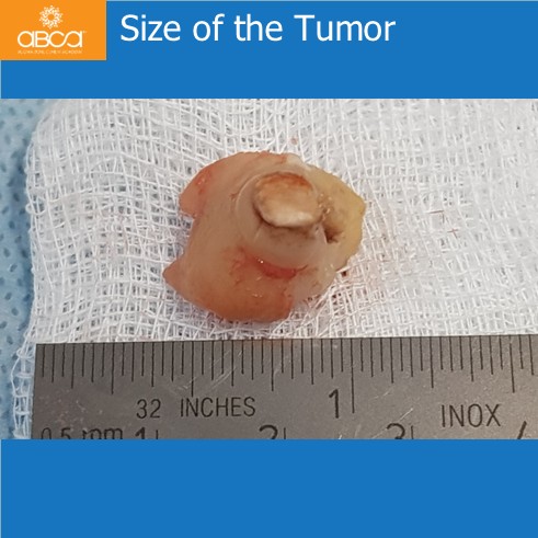
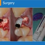
A 32-year-old male patient presents with a long-standing problem in the left mandibular region. Previous clinicians did not attempt removal of the lesion. Diagnoses of the lesion is a complex odontoma fused with tooth #34 (21), with destruction of tooth #35 (20). CBCT imaging reveals involvement of both cortical plates in this area.
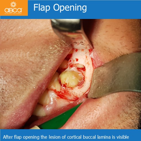
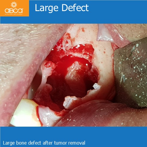
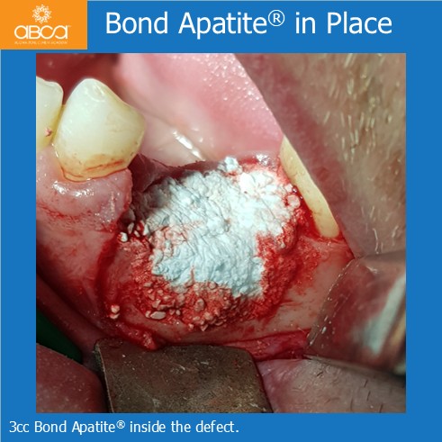
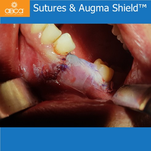
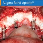
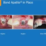
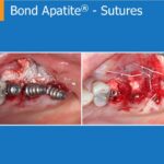
Tumor removal with peripheral bone curettage to a depth of approximately 1 mm and extracted teeth #34 (21) and #35 (20). Augmentation of the bone defect with 3 cc of Bond Apatite® and coverage with Augma Shield™, secured to the wound using Periacryl 90 adhesive. Histopathological examination done on the excised tumor confirms the diagnosis of complex odontoma.
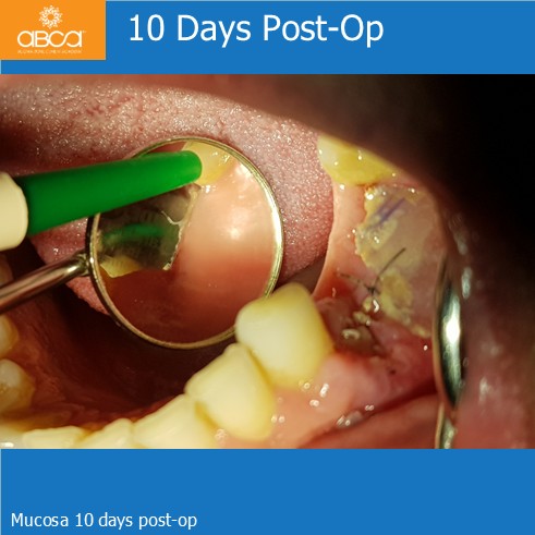
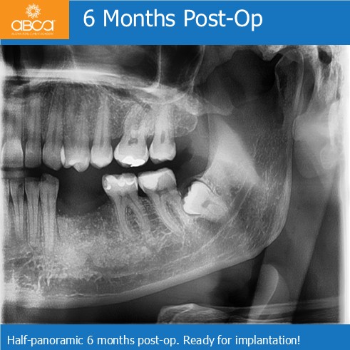
The wound healed well, suture removal takes places 10 days post-op. Six months follow-up evaluation demonstrates excellent regeneration of the bone defect. The newly formed bone is now suitable for implant placement.